
When it comes to spotting problems in the back of the eye, doctors don’t just rely on asking you if your vision is blurry. They use powerful imaging tools that show details you can’t see with your own eyes. Three of the most important tools are OCT, fundus photography, and angiography. Each one gives a different kind of picture - like taking X-rays, snapshots, and video of your retina all at once. Together, they help catch diseases early, track how they change over time, and decide what treatment you need.
What OCT Actually Shows
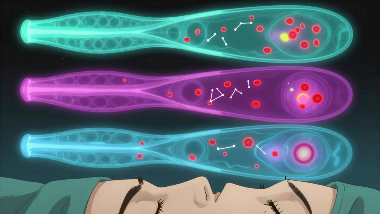
Optical Coherence Tomography, or OCT, doesn’t use light like a camera. It uses sound waves made of light - called interferometry - to build cross-sections of your retina, layer by layer. Think of it like slicing an onion, but instead of peeling it apart, you’re seeing each layer in perfect detail on a screen.
Modern spectral-domain OCT (SD-OCT) can show structures as thin as 5 to 7 micrometers. That’s about 1/10th the width of a human hair. It catches tiny changes in the retina before you even notice vision loss. For example, if you have early macular degeneration, OCT can spot fluid buildup under the retina long before your vision gets blurry. In diabetic eye disease, it shows swelling in the macula, even when it’s too mild to affect sight.
There’s also swept-source OCT (SS-OCT), which scans faster - up to 400,000 lines per second - and goes deeper. It’s especially good at showing the choroid, the layer beneath the retina that supplies blood. This matters because some diseases, like punctate inner choroidopathy, start there. SS-OCT can reveal inflammation or thinning in that layer, which older OCT machines might miss entirely.
Fundus Photography: The Snapshot of Your Retina
Fundus photography is the oldest of the three, but it’s still essential. It’s basically a high-res photo of the back of your eye - the retina, optic nerve, and blood vessels. Cameras like the Zeiss FF 450+ capture these images in seconds. They’re used to document changes over time. If your doctor sees a new spot on your retina, they take a photo. Next year, they compare it. If it’s grown, they know the disease is active.
This method is great for spotting diabetic retinopathy, retinal hemorrhages, or signs of glaucoma. But it has limits. It can’t show fluid inside the layers of the retina. It can’t tell if a blood vessel is leaking. It’s like taking a picture of a car’s exterior - you can see scratches and dents, but you can’t see if the engine is failing.
Fluorescein Angiography: Tracking Blood Flow in Real Time
Fluorescein angiography (FA) is where things get dramatic. A fluorescent dye is injected into your arm, and as it travels through your bloodstream, a camera takes rapid photos of your retina. The dye lights up the blood vessels, letting doctors see exactly where blood is leaking, pooling, or not flowing at all.
This is the gold standard for detecting leakage in diabetic macular edema. One study found FA was 100% sensitive at spotting leakage, while OCT only caught 79%. That 21% gap? It’s the difference between missing a problem and catching it early enough to prevent vision loss.
But FA has downsides. It’s invasive. The dye can cause nausea, vomiting, or, rarely, an allergic reaction. The whole process takes 10 to 30 minutes. You’ll have yellow-tinted skin and urine for hours. And it can’t show the deeper layers of the retina - just the surface vessels.

OCT Angiography: The Non-Invasive Revolution
Enter OCT angiography (OCTA). This is the newest player, and it’s changing how eye doctors work. OCTA uses the same machine as regular OCT but adds software that detects moving blood cells. No dye. No injection. Just a quick scan - sometimes under 10 seconds.
OCTA can separate the retina’s blood vessels into three distinct layers: superficial, middle, and deep. That’s huge. In diabetic retinopathy, early damage happens in the deep capillaries. FA might miss it. OCTA shows it clearly. Studies show OCTA detects more microaneurysms and non-perfused areas than FA, especially in the peripheral retina.
One study found that OCTA spotted 57% of retinal capillary hemangiomas that SD-OCT missed. Another showed it revealed hidden areas of poor blood flow in patients with punctate inner choroidopathy - areas that didn’t show up on any other test.
But OCTA isn’t perfect. It can’t show dye leakage. If a vessel is leaking fluid, OCTA won’t see it. That’s why FA still has a place. Also, if you move your eye during the scan - even slightly - the image gets blurry. That’s hard for older patients or those with nystagmus.
How These Tools Work Together
No single tool gives the full picture. That’s why doctors use them in combination. Here’s how they fit together:
- For macular holes or swelling: OCT is king. It shows the exact depth and size of the problem.
- For diabetic retinopathy: Use OCT to check for fluid, FA to check for leakage, and OCTA to see if capillaries are dying off.
- For Coats disease: OCT finds hidden fluid pockets and cholesterol crystals under the retina - things photos can’t show. FA reveals abnormal blood vessels. Together, they confirm the diagnosis.
- For rare conditions like PIC: OCTA shows tiny areas of choriocapillaris blockage. FA and indocyanine green angiography help confirm it’s not another disease.
A 2023 study in the MJCU Journal showed that combining all four - OCT, fundus photos, FA, and OCTA - gave the clearest picture of disease progression. In fact, some lesions were only visible when all four were reviewed together.
What’s Changing Now
The biggest shift is speed and depth. Newer OCTA machines, like the Spectralis platform, now offer wider fields of view. That means they can capture more of the peripheral retina in one scan. This is critical because early signs of disease often appear far out in the periphery - areas older machines couldn’t reach.
Also, algorithms are getting smarter. Software can now automatically measure the size of the foveal avascular zone (FAZ) - the tiny area in the center of the retina with no blood vessels. If it grows, it’s a warning sign. Some systems even flag areas of non-perfusion and suggest possible diagnoses.
But training hasn’t caught up. Most ophthalmologists learned to read FA and OCT. OCTA is different. It’s not just about seeing vessels - it’s about spotting artifacts that look like vessels. A shadow from a blood vessel can mimic a blockage. A blink can look like a capillary dropout. That’s why specialists now take extra training just to interpret OCTA correctly.
What You Should Know as a Patient
If you’re being tested for a retinal disease, you might go through all three - or just two. Here’s what to expect:
- OCT: You’ll sit in front of a machine, rest your chin, and stare at a light. No contact. No discomfort. Takes 5 minutes.
- Fundus photo: A bright flash. It might surprise you, but it’s quick. No pain.
- Fluorescein angiography: A needle in your arm. You’ll feel a warm flush. Your skin might turn yellow. Wait 30 minutes. Bring someone to drive you home.
- OCTA: Same as OCT. No injection. Just sit still. It’s faster than the others.
The biggest benefit? OCTA eliminates the risks of dye. No more nausea. No more allergic reactions. For patients who’ve had bad reactions before, this is a game-changer.
But don’t assume OCTA replaces everything. If your doctor says you still need FA, it’s because they need to see leakage - something OCTA can’t show.
What’s Next
The future of eye imaging is automatic. AI is being trained to spot early signs of macular degeneration or diabetic damage in OCT scans - faster than any human. Some systems now predict disease progression based on changes over time. In five years, you might get a report that says, “Your deep capillary layer has lost 12% of blood flow since last year. Risk of vision loss: moderate.”
For now, the best tool is still the one used together. OCT shows structure. FA shows leaks. OCTA shows blood flow. Fundus photos show the big picture. Used right, they’re not just images - they’re early warnings that can save your vision.
I read this whole thing. Honestly? Too much jargon. I just want to know if I need to freak out about my yearly eye scan. You guys make it sound like my retina is a warzone. Can we just say 'get checked' and leave it at that?
This is actually one of the clearest breakdowns I've seen. It's not just about tech-it's about preserving something irreplaceable: your ability to see your grandkids' faces. Every scan, every detail, matters.
OCTA is a game changer. No dye, no nausea, just quick scans. My mom had a bad reaction to FA last year. She cried. Now she actually looks forward to her eye appointments. That’s huge.
Let’s be real. These ‘non-invasive’ tools are just corporate marketing. The real money is in repeat scans. They’ll keep pushing OCTA because it’s cheaper than FA, not because it’s better. You think they care if you miss 21% of leakage? They care if you come back next year.
I love how you explained the layers. It made me feel like my eyes aren’t just a blob of tissue-they’re this intricate, living map. I’ve had diabetic retinopathy for 8 years, and this is the first time I actually feel like I understand what’s happening. Thank you.
The article mentions AI predicting progression. That’s terrifying. Who’s training these algorithms? What data are they using? What if they’re biased toward certain ethnicities? We’re automating diagnosis without asking who’s at risk of being misdiagnosed.
OCTA is like magic. Imagine your retina as a city, and the blood vessels are highways. OCTA doesn’t just show traffic-it shows which lanes are crumbling, which exits are blocked, and where the construction crews are hiding. It’s not just imaging. It’s urban planning for your vision.
We treat the eye like a camera, but it’s not. It’s a living organ that talks in whispers. These tools? They’re translators. And we’re just learning the language. Maybe the real breakthrough isn’t the tech-it’s learning to listen.
All this tech, and yet 40% of diabetic patients still lose vision. Why? Because they don’t get screened. The real issue isn’t imaging-it’s access. You need insurance, transportation, time off work. This article reads like a luxury catalog. For most people, ‘get an OCT’ is a fantasy.