
When it comes to spotting problems in the back of your eye, doctors don’t just rely on a light and a magnifying glass anymore. Modern eye care depends heavily on three key imaging tools: OCT, fundus photography, and fluorescein angiography. These aren’t just fancy gadgets-they’re the reason many people keep their vision longer, even with serious conditions like diabetes or macular degeneration. Each one shows something different, and together, they give a full picture of what’s happening inside your retina.
What OCT Does: Seeing Layers You Can’t See
Optical Coherence Tomography, or OCT, is like an ultrasound for your eye-but it uses light instead of sound. It takes detailed cross-sections of the retina, showing each layer like slices of bread. This lets doctors see swelling, thinning, or holes in the retina that aren’t visible during a regular exam.
Modern spectral-domain OCT (SD-OCT) can resolve details as small as 5 to 7 micrometers. That’s about one-tenth the width of a human hair. It’s especially good at catching early signs of macular edema, where fluid leaks into the macula and blurs central vision. It also spots macular holes, epiretinal membranes, and changes in the optic nerve from glaucoma.
There’s also swept-source OCT (SS-OCT), which goes deeper and scans faster-up to 400,000 scans per second. That means it can see the choroid, the layer under the retina that supplies blood. This is critical for conditions like choroidal neovascularization, where abnormal blood vessels grow and leak under the retina. SS-OCT is now becoming the new standard in busy clinics because it’s faster, clearer, and works better through cloudy lenses or small pupils.
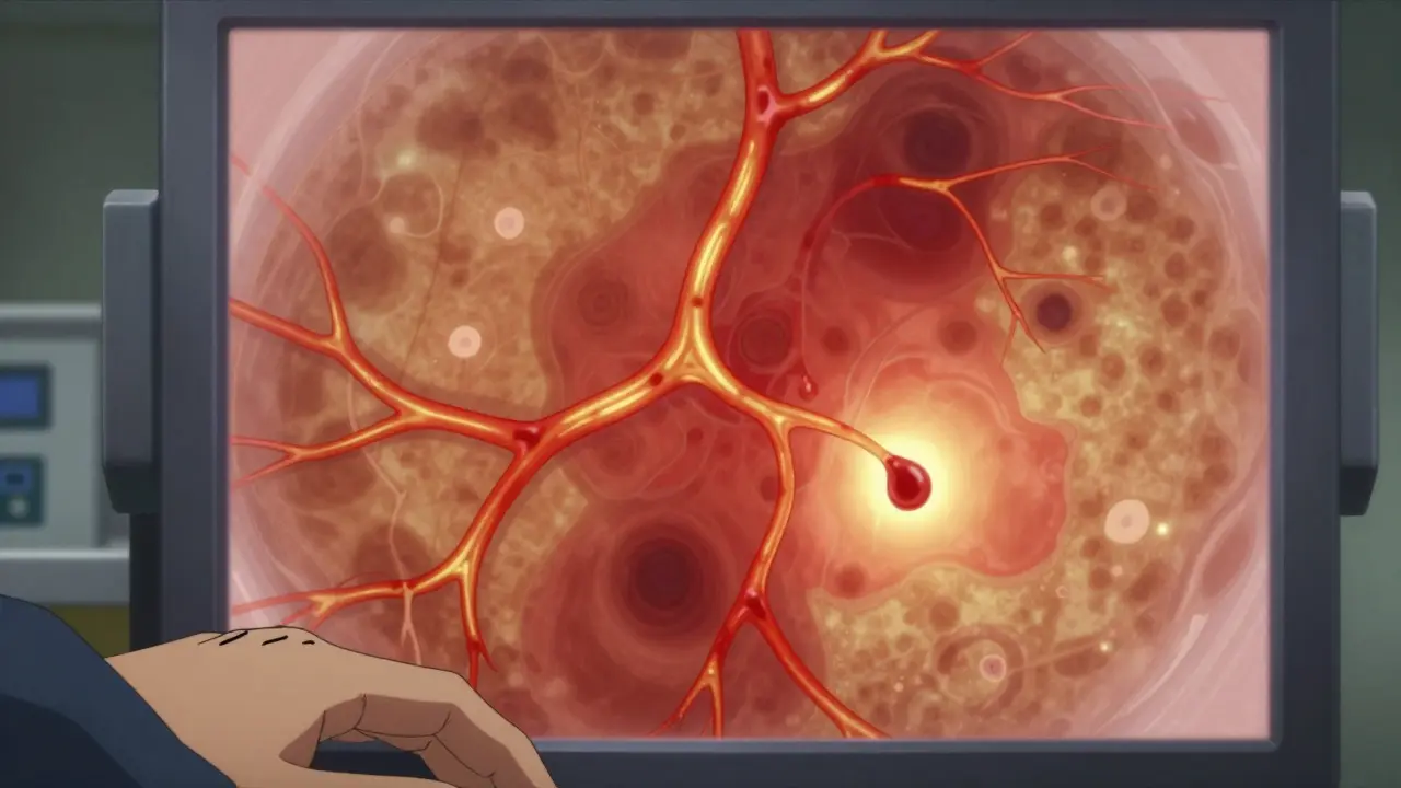
Fundus Photography: The Snapshot of Your Retina
Fundus photography is exactly what it sounds like: a high-res photo of the back of your eye. Special cameras like the Zeiss FF 450+ capture the optic disc, macula, blood vessels, and retina in vivid color. It’s the go-to tool for documenting changes over time.
For diabetic retinopathy, this is essential. Doctors compare photos year after year to see if new blood vessels are forming or if there’s bleeding in the retina. It’s also used to track age-related macular degeneration, retinal detachments, and even rare diseases like Coats disease or punctate inner choroidopathy.
But fundus photos have limits. They show surface details, not depth. A swollen retina might look normal in a photo if the swelling is subtle. That’s why they’re almost always paired with OCT. A photo tells you where something is wrong; OCT tells you how much and why.
Fluorescein Angiography: Tracking Blood Flow in Real Time
Fluorescein angiography (FA) has been around since the 1960s, and it still holds its place. It works by injecting a yellow dye into a vein in your arm. As the dye travels through the bloodstream, a camera takes a rapid series of photos to show how the dye moves through the retinal blood vessels.
This is the only way to see exactly where blood vessels are leaking, blocked, or growing abnormally. For diabetic macular edema, FA is more sensitive than OCT at detecting subtle leakage. One study found that FA caught leaks in 100% of cases where OCT only saw them in 79%. That 21% gap matters-it can mean the difference between timely treatment and permanent vision loss.
But FA isn’t perfect. It’s invasive. The dye can cause nausea, vomiting, or, rarely, allergic reactions. It takes 10 to 30 minutes to complete. And because it’s based on dye movement, it’s subjective. Two doctors might interpret the same image differently.
OCT Angiography: The Non-Invasive Breakthrough
Enter OCT angiography (OCTA). This is OCT’s smarter, dye-free cousin. Instead of injecting dye, OCTA detects tiny movements in red blood cells to map blood flow. It creates 3D images of the capillaries in the retina and choroid-without a needle.
It’s fast. A full scan takes seconds. It’s safe. No dye means no side effects. And it can separate the retina into three layers: superficial, middle, and deep capillary plexuses. This lets doctors see exactly where blood flow is failing.
Studies show OCTA is better than FA at spotting early signs of proliferative diabetic retinopathy, especially tiny new blood vessels near the optic nerve. In one study of 84 eyes with retinal capillary hemangiomas, SS-OCTA found 57% that SD-OCT missed. It’s also better at detecting non-perfusion areas in diseases like punctate inner choroidopathy, where tiny patches of dead blood vessels are invisible on traditional imaging.
But OCTA has blind spots. It can’t see leakage the way FA does. If fluid is seeping out of a vessel but not pooling, OCTA might not catch it. It’s also sensitive to eye movement. If you blink or shift your gaze, the image gets blurry. That’s why it’s not a full replacement for FA-yet.
How They Work Together
No single tool tells the whole story. That’s why experts use them together.
- For diabetic retinopathy: Fundus photos track long-term damage, OCT measures swelling, and FA spots leakage. OCTA adds detail on new blood vessel growth without the risk of dye.
- For macular degeneration: OCT shows fluid under the retina; FA confirms if it’s from abnormal vessels; OCTA maps the exact pattern of those vessels.
- For Coats disease: Fundus photos show the whitish exudates; OCT reveals fluid pockets and cholesterol crystals hidden beneath; FA confirms vessel abnormalities.
- For punctate inner choroidopathy: OCTA is often the first to show choroidal non-perfusion, while FA and fundus autofluorescence help confirm the diagnosis.
Doctors now rely on what’s called multimodal imaging. Think of it like a team: fundus photos are the wide-angle view, OCT is the MRI, and FA/OCTA are the blood flow tracers. Together, they catch problems earlier and track treatment more accurately.

What’s Changing Now
The biggest shift in the last five years is the rise of swept-source OCT and wide-field OCTA. Systems like the Spectralis OCTA can now scan a much larger area of the retina in one go-up to 120 degrees. That means they can see problems near the edge of the retina, where FA used to be the only option.
Artificial intelligence is starting to help too. Some systems now auto-detect fluid, measure vessel density, or flag areas of non-perfusion. This reduces human error and makes tracking changes over time more precise.
But the biggest win for patients? Less discomfort. No more waiting 20 minutes after a dye injection. No more nausea. No more blurry vision for hours. OCTA lets people get the same-or better-information without the hassle.
Limitations and What to Expect
None of these tools are magic. OCTA can’t replace FA in all cases. FA still wins when you need to see fine leakage patterns. OCT can’t see through dense cataracts or vitreous hemorrhage. Fundus photos can miss early swelling.
Also, not every clinic has all three. Smaller practices might only have OCT and fundus cameras. That’s okay-they’ll refer you out if they need angiography. The goal isn’t to have every tool; it’s to use the right one at the right time.
For patients, the experience is simple: you sit in front of a machine, stare at a light, and hold still. No pain. No needles (unless FA is needed). Most scans take less than 10 minutes total. Results come back quickly, and doctors can show you the images on screen right away.
What You Should Know
If you have diabetes, glaucoma, macular degeneration, or a family history of retinal disease, these imaging tests aren’t optional-they’re preventive. Catching a problem early means treatment can stop vision loss before it starts.
Don’t be afraid to ask your doctor: “Which imaging tests are you using, and why?” Understanding what each one shows helps you make better decisions. And if your doctor only uses one type of imaging, ask if adding another could give a clearer picture.
The future of eye care isn’t about replacing tools-it’s about combining them. OCT, fundus photos, and angiography each have strengths. Together, they’re giving people with eye disease a real shot at keeping their sight.
Is OCT the same as a retinal scan?
Yes, OCT is a type of retinal scan-but it’s more detailed than older methods. While traditional retinal scans take a flat photo of the back of the eye, OCT creates 3D cross-sections that show individual layers of the retina. Think of it like going from a photograph to an MRI.
Do I need dye for OCT angiography?
No. OCT angiography (OCTA) uses light to detect blood flow, so no dye is injected. This makes it safer and faster than traditional fluorescein angiography. It’s ideal for people who are allergic to dye, pregnant, or just want to avoid needles.
Why would my doctor order both OCT and fluorescein angiography?
Because they show different things. OCT shows structure-like swelling or holes in the retina. Fluorescein angiography shows function-where blood is leaking or blocked. For conditions like diabetic macular edema or retinal vein occlusion, you need both to get the full picture. One can’t replace the other yet.
Can OCT detect glaucoma?
Yes. OCT measures the thickness of the retinal nerve fiber layer, which thins as glaucoma damages the optic nerve. It’s one of the most reliable tools for early detection, often spotting changes years before vision loss becomes noticeable.
How often should I get these scans?
It depends on your condition. For healthy people, no routine scans are needed. If you have diabetes, annual OCT and fundus photos are standard. For macular degeneration or glaucoma, scans every 3 to 6 months are common. Your doctor will tailor the schedule based on your risk and how your disease is progressing.
It's wild how far we've come from just poking around with a light and a magnifying glass. I remember my grandpa complaining about eye exams back in the '80s-he’d get his pupils dilated, sit in a dark room for an hour, and still leave with more questions than answers. Now? You sit there, stare at a light, and ten seconds later the doctor’s showing you a 3D model of your retina like it’s a Google Earth zoom-in. No more guessing. No more "maybe it’s just aging." If your macula’s got a tiny hole, they’ll show it to you in color. It’s like having an MRI for your eyeball.
And honestly, the fact that we can now map capillary flow without dye? That’s huge. I’ve had FA done before. The nausea, the yellow tint in your vision for hours, the whole "why did I agree to this" vibe. OCTA feels like a gift. No needles, no waiting, no "oh god I think I’m gonna puke." It’s not perfect, sure, but for routine monitoring? It’s a game-changer.
Still, I worry about over-reliance. Some clinics are ditching FA entirely because OCTA is "easier." But I’ve seen cases where the leakage was so subtle, OCTA missed it completely. FA still catches the ghost leaks-the ones that don’t pool but still creep. You can’t replace a functional map with a structural one. It’s like trying to diagnose a leaky pipe by looking at the walls instead of turning on the water.
And don’t get me started on AI. Yeah, it flags fluid and vessel density like a boss, but I’ve seen it miss a small neovascularization because the algorithm was trained on white retinas. What about darker skin tones? What about patients with cataracts that distort the scan? AI doesn’t ask questions. It just spits out a number. Human eyes still matter.
Bottom line: we’ve got tools now that can catch problems before you even notice blurry vision. That’s incredible. But the best diagnosis still comes from a doctor who knows when to trust the machine-and when to trust their gut.
o dont get me started on octa i had a scan last year and the tech said "no abnormalities" but then my doc pulled up the fa and said "oh wow theres a leak right here" turns out the octa missed it because i blinked. so yeah its cool but dont trust it blindly
octa is just a marketing gimmick pushed by big ophthalmology to sell more machines. fa has been reliable for 60 years and now they want you to pay extra for a scan that cant even see leakage. theyre just trying to upsell. and dont even get me started on ai interpreting images. what happens when it misses something and you go blind? who gets sued? the company that made the algorithm?
One cannot help but observe that the evolution of retinal imaging represents not merely a technological advancement, but a profound epistemological shift in the phenomenology of medical perception. Where once the physician relied upon the corporeal gaze-a tactile, embodied, and inherently fallible act of observation-we now defer to algorithmic renderings of biological structure. The retina, once a canvas of organic complexity, has been reduced to a dataset: layers digitized, vessels quantified, perfusion mapped as spectral gradients.
Yet, in this digitization, we risk a quiet alienation from the patient’s lived experience. The clinical encounter, once a dialogue of symptoms, history, and intuition, is increasingly mediated by screens, metrics, and automated classifications. The patient no longer describes blurred vision-they point to a red zone on a heatmap. The doctor no longer asks, "Have you noticed changes?"-they query, "Does this vessel density fall below the 5th percentile?"
Is this progress? Or merely the colonization of medicine by the logic of efficiency? The dye-free OCTA is elegant, yes. But elegance without epistemic humility is dangerous. We must remember: the machine sees patterns. The healer sees persons.
I love how this article breaks down the tools without jargon. I work in a community clinic and we only have OCT and fundus photos, but we’ve been able to catch so many early cases of diabetic retinopathy just by comparing yearly images. One woman came in thinking her vision was just getting "fuzzy with age"-turns out her retina had early swelling. We referred her, she got treatment, and now she’s driving again. That’s the real win.
Also, I’ve had patients who were terrified of FA because of the dye. When I explain OCTA is just a quick scan with no needles, they relax. It’s not perfect, but it removes a huge barrier. More people get screened. More people keep their sight. That’s what matters.
And yeah, AI is still learning. But I’ve seen it catch a tiny hemorrhage I missed. Not because it’s smarter, but because it doesn’t get tired. We need both.
so what if the government is using these scans to track your eye health and then sell your data to insurance companies? they already know if you have diabetes or glaucoma. what if they raise your premiums because your retina shows "early signs"? theyre not doing this for your health. theyre doing this to control you. i read online that some clinics are already sharing scan data with health insurers. dont let them fool you.
Just a heads up-some of you are acting like OCTA is magic. It’s not. I’m an optometrist. We use all three tools daily. OCTA is amazing for spotting early capillary dropout in diabetic eyes, but if you have vitreous hemorrhage? It’s useless. The image just turns to static. FA still wins there. And fundus photos? They’re the only way to catch peripheral exudates in Coats disease. You need the whole team.
Also, stop blaming AI. It’s not the algorithm that missed the leak. It’s the tech who didn’t re-scan after the patient blinked. Human error is still the #1 cause of misdiagnosis. Tools help. They don’t replace.
And for the conspiracy folks-no, your retina scan isn’t being sold to insurers. The data is HIPAA-protected. If you’re worried, ask your doc. They’ll show you the consent form. But if you skip the scan? That’s when you lose vision. Not because of the government. Because you didn’t show up.
I’ve had glaucoma for 8 years. Every 6 months I get an OCT. I used to hate it. Felt like they were poking around in my soul. But now? I look forward to it. The doctor shows me the nerve layer thickness on screen. Last year it was 82 microns. This year? 81.5. That’s a tiny drop. But over time? It adds up. I know when to push for treatment before I lose more vision.
And honestly? The fact that I can see it myself-that I’m not just trusting someone else’s word-that’s empowering. I’m not a patient. I’m a participant.
Don’t let fear stop you from getting scanned. The worst thing isn’t the machine. It’s waiting too long.
yo i got my first OCTA last week and it was wild. no dye, no waiting, just stare at a light and boom-your eye’s blood vessels are on screen like a subway map. my doc pointed out a dead zone near my optic nerve i didn’t even know existed. turns out i’ve had early signs of non-perfusion since 2021. i’m lucky i caught it. now i’m on a treatment plan. if i hadn’t gotten scanned? i’d be blind by now. so yeah, trust the tech. it saved my sight.
biggest thing people dont get is that these tools arent about scaring you. theyre about giving you power. if you know your retina is thinning, you can change your diet, control your sugar, quit smoking, get more sleep. you’re not just waiting for blindness. you’re fighting it. and that changes everything.
also, if your doc only uses one tool? ask for more. not because you’re paranoid. because you care. your eyes are worth it.
There’s something deeply human about seeing your own retina on a screen. Not as a diagnosis, but as a map of your life-the years of sugar, the stress, the sleepless nights. It’s not just a medical tool. It’s a mirror. And when you see the damage, you’re not just a patient. You’re a person who can choose to change.
I’ve watched patients cry-not from fear, but from relief. "I didn’t realize it was this bad." Then they take action. That’s the quiet power of imaging. It doesn’t just show disease. It shows possibility.
Let me be perfectly clear: the use of OCT, FA, and OCTA is not merely a clinical procedure-it is a systemic surveillance mechanism. The manufacturers of these devices are owned by conglomerates with ties to pharmaceutical giants, defense contractors, and data brokers. The very act of scanning your retina generates a biometric signature that can be stored, sold, and weaponized. Have you considered that your retinal vascular pattern is as unique as your fingerprint? And that this data is being aggregated into national health databases without informed consent? I have reviewed the FDA’s 510(k) submissions. The disclosures are deliberately vague. This is not medicine. This is social engineering.
And don’t forget: the dye used in FA contains fluorophores that may accumulate in neural tissue. Long-term effects? Unknown. But the FDA has approved it under "substantial equivalence." That’s not safety. That’s negligence. Demand transparency. Demand autonomy. Demand truth.
so you're telling me i can get a full eye scan without getting injected with dye? and it's faster than my coffee break? sign me up. also, why does my doctor charge $400 for this? i thought this was supposed to be healthcare, not a luxury spa for eyeballs.
My mom has macular degeneration. We used to do FA every 3 months. She hated it. Nausea, blurry vision for hours, crying in the waiting room. Last year we switched to OCTA. She didn’t even notice it happened. Just sat there, stared at a light, and walked out. Same info. No trauma. That’s not just better tech. That’s dignity.
If you’re scared of scans? Don’t be scared of the machine. Be scared of waiting. One early scan can save your sight. Don’t let fear cost you your world.
FA still matters. OCTA can’t see leakage. I’ve seen it. If your doctor skips FA for diabetic edema, they’re missing half the picture. Don’t assume one tool does it all. Ask for both. Your vision is worth it.